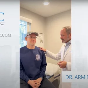


Under Dr. Tehrany’s guidance, Manhattan Orthopedic Care provides arthroscopic chondroplasty—a minimally invasive procedure that smooths and repairs damaged cartilage to relieve pain, enhance mobility, and maintain joint health for improved patient results in New York City and Staten Island.
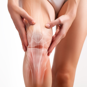
What Is Chondroplasty of the Knee?
Arthroscopic chondroplasty is a minimally invasive surgical approach used to address cartilage damage within a joint, typically the knee. Using a slender camera known as an arthroscope, Dr. Tehrany can visualize the damaged cartilage on a monitor, and then employ specialized instruments inserted through tiny incisions to remove loose fragments and smooth rough surfaces. This process helps reduce pain, restore mobility, and potentially slow the progression of joint deterioration. Because it is performed arthroscopically, patients usually experience less post-operative discomfort and shorter recovery times compared to open surgery. At Manhattan Orthopedic Care, we utilize advanced techniques to provide personalized treatment aimed at preserving joint function and enhancing quality of life. Following the procedure, patients will undergo a rehabilitation plan to optimize healing and maximize long-term results.
Conditions Treated With Arthroscopic Chondroplasty
Arthroscopic chondroplasty helps address a variety of conditions where cartilage damage hinders normal joint function. From everyday wear and tear to acute injuries, this minimally invasive procedure can relieve pain, restore mobility, and slow further deterioration, making it a valuable treatment for individuals seeking long-term joint health and comfort.
Osteoarthritis
Osteoarthritis occurs when cartilage thins from prolonged wear, causing joint stiffness and discomfort. Arthroscopic chondroplasty helps by removing loose debris and smoothing rough surfaces, reducing pain and restoring proper movement.
Chondromalacia Patella
Chondromalacia patella involves softening of the cartilage under the kneecap, leading to pain and grinding sensations. Patella Chondroplasty smooths these surfaces, alleviating discomfort and allowing more efficient kneecap movement and alignment.
Cartilage Tears from Sports Injuries
Sports-related impacts or twisting motions can tear cartilage, causing pain and swelling. During arthroscopic chondroplasty, surgeons trim ragged edges and smooth injured areas, helping restore stability and preventing further deterioration.
Degenerative Joint Changes
Degenerative joint changes emerge from aging, repetitive stress, or genetic factors, wearing away protective cartilage. Chondroplasty targets damaged sites to mitigate discomfort, promote smoother movement, and delay extensive joint interventions.
See Arthroscopic Knee Chondroplasty in Action
Check out our video gallery to discover how Dr. Tehrany can restore your joint function and comfort!
Manhattan Orthopedic’s Advanced Approach to Arthroscopic Chondroplasty
Manhattan Orthopedic Care stands out for its expertise in advanced arthroscopic procedures, ensuring personalized care for each patient. Dr. Tehrany’s unparalleled commitment to minimally invasive surgery, combined with his compassionate approach, has made him a sought-after orthopedic specialist in New York City. From the initial assessment to recovery, the dedicated team emphasizes patient education, comfort, and long-term joint health. With innovative techniques and a patient-centered philosophy, Manhattan Orthopedic Care is your trusted partner for effective chondroplasty solutions.
- Dr. Tehrany is board-certified by the American Board of Orthopaedic Surgery
- Manhattan Orthopedic Care’s welcoming environment fosters open communication and personalized treatment plans. Every patient is guided through each step, ensuring clear expectations, compassionate support, and an overall positive surgical experience.
- With cutting-edge equipment and advanced techniques, Dr. Tehrany achieves minimal downtime and rapid healing. Patients benefit from effective treatment that reduces discomfort, preserves healthy cartilage, and safeguards long-term joint functionality.
Your Arthroscopic Chondroplasty Staten Island Surgery
This outpatient procedure is used to repair a small area of damaged cartilage in the knee. The damaged tissue is removed, allowing healthy cartilage to grow in its place. It is performed through small incisions on the sides of the knee with the aid of a small video camera called an arthroscope.
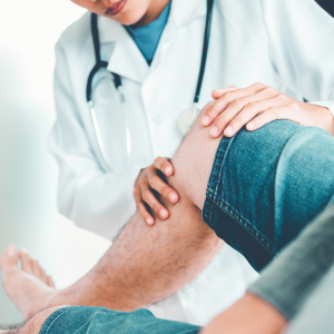
Initial Arthroscopic Chondroplasty Consultation
During your initial arthroscopic chondroplasty consultation, Dr. Tehrany will examine your joint, discuss your symptoms and medical history, and determine the best approach for optimal treatment.
Pre-surgery preparation
Before surgery, you’ll receive detailed instructions on tests, medication adjustments, and any lifestyle changes needed. Arrive well-rested, follow fasting guidelines, and stay hydrated for a smoother surgical experience.
Day of Surgery
- Preparation for arthroscopic knee chondroplastyThe patient is positioned so that the knee is clearly visible to the physician, and the area is cleaned and sterilized.
- Accessing the knee jointThe surgeon creates two to five small incisions around the front of the knee. An arthroscopic camera is inserted through one of the incisions. The other incisions will be used as access points for other arthroscopic tools.
- Surveying the jointFluid is pumped in to expand the joint, giving the surgeon a clear view and room to work. The surgeon uses the arthroscope to identify the problem area.
- Removing the damaged cartilageThe surgeon inserts small surgical tools and carefully removes the small patch of damaged cartilage and any loose tissue.
- End of the knee surgery and knee recovery
The excess fluid is drained from the knee, the instruments are removed and the incisions are closed. As the knee heals, new “scar tissue” cartilage will grow over the bare spot to replace the missing cartilage.
Follow-up Appointment
Regular follow-up appointments allow Dr. Tehrany to evaluate healing progress, address concerns, and adjust your rehabilitation plan. Consistent communication ensures a safe recovery and long-term joint function.
Schedule Your Surgery Consultation
Ideal Candidates for Arthroscopic Chondroplasty in New York
Arthroscopic chondroplasty in New York is recommended for patients experiencing cartilage damage who desire minimally invasive solutions to relieve pain and restore function.
- Healthy enough for a minimally invasive surgical procedure
- Suffering from cartilage tears, wear-and-tear, or localized joint damage
- Seeking pain relief and improved mobility in affected joints
- Non-responsive to conservative treatments like physical therapy or medications
- Willing to follow postoperative rehabilitation for long-term success
Your Recovery Journey from Arthroscopic Knee Chondroplasty
Recovery timelines after arthroscopic chondroplasty vary based on factors like overall health and the extent of cartilage damage. Patients can expect initial swelling and mild discomfort, which typically subsides with rest, ice, and prescribed pain management. A supportive brace or crutches may be recommended to limit weight-bearing during the early stages. Physical therapy plays a vital role in regaining mobility and strengthening the surrounding muscles, so adhering to a personalized rehabilitation plan is essential. Over time, most individuals experience noticeable pain relief and improved joint function, enabling them to gradually return to regular activities and an active lifestyle.
Benefits of Arthroscopic Chondroplasty
Arthroscopic chondroplasty delivers minimally invasive relief and effectively helps preserve cartilage, improving joint function, alleviating pain, and supporting long-term mobility.
- Minimally invasive approach reduces postoperative pain and scarring
- Helps maintain stable, functioning cartilage for joint support
- Enhances overall mobility and range of motion
- May delay or prevent more invasive procedures
- Supports quicker recovery with targeted therapy
Understanding the Potential Risks of Arthroscopic Chondroplasty in Staten Island
While arthroscopic chondroplasty is generally safe, potential complications can occur. With Dr. Tehrany’s expertise, these risks are significantly minimized, but may include infection, blood clots, or nerve and vessel damage. Knowing them helps ensure an informed approach and proper aftercare.
- Infection at the incision site
- Blood clots from restricted mobility
- Nerve or blood vessel damage
- Incomplete relief of pain symptoms
- Possible joint stiffness during healing
Other Knee Surgery Options Offered At Manhattan Orthopedic Care, NY
Manhattan Orthopedic Care offers a broad spectrum of knee procedures, from minimally invasive arthroscopic repairs to full surgical reconstructions, ensuring treatments that restore stability, relieve pain, and improve joint function.
ACL Reconstruction
ACL reconstruction is performed arthroscopically, making it a minimally invasive procedure. The procedure replaces a torn ligament with a graft to restore knee stability, significantly enhance mobility, and promote rapid, lasting recovery.
Meniscus Repair
Meniscus repair employs arthroscopic techniques to mend torn knee cartilage. This minimally invasive procedure preserves natural cushioning, reduces pain, and prevents further joint degeneration, ultimately promoting healing and enhanced mobility.
Knee Arthroscopy
Knee arthroscopy uses small incisions to diagnose and treat internal joint issues. This minimally invasive procedure facilitates precise interventions, shortens recovery time, and minimizes the need for full knee surgery.
Book Your Consultation Today!
Take the first step toward a pain-free life. Schedule your consultation now to explore personalized knee treatments designed for rapid recovery, lasting stability, and improved mobility.
Schedule a Shoulder Consultation
Read More About Arthroscopic Chondroplasty in New York
FAQ
What type of anesthesia is used during arthroscopic knee chondroplasty, and how long does the procedure take?
Arthroscopic chondroplasty is performed under general or regional anesthesia. Typically, the procedure lasts approximately 45 minutes to an hour, depending on cartilage damage complexity.
Are there non-surgical alternatives to arthroscopic chondroplasty in Staten Island?
Non-surgical options, such as physical therapy, medications, and injections, may help manage symptoms but typically do not repair cartilage damage as effectively as arthroscopic chondroplasty. Contact Dr. Tehrany to determine the optimal procedure for your needs.
When can I safely resume physical activities or sports following the procedure?
Patients often begin gentle range-of-motion exercises 2-3 weeks post-surgery, yet full sports participation may require 2-3 months of rehabilitation for optimal recovery and safe return to high-impact activities.